Research
OB-GYN
PELVIC FLOOR SCANNING
Background
The female pelvic floor (PF) muscles provide support to the pelvic organs. During delivery, some of these muscles have to stretch up to three times their original length to allow for the passage of the baby, leading frequently to damage and consequently later life PF dysfunctions. Three-dimensional (3D) ultrasound (US) imaging can be used to image these muscles and to diagnose these damages by assessing quantitative, geometrical and functional information of the muscles through strain imaging. In our work we have developed 3D US strain imaging of the PF muscles and explored its application on the puborectalis muscle (PRM), which is one of the major PF muscles. Our work is funded by the GYNIUS grant from NWO.
Methods
In our work, US data from female pelvic floor is obtained by the radiologist through transperineal US. Data acquisition takes place while the muscle voluntarily deformed, for example, from rest to contraction. The 3D US volumes are then used to measure the displacement and deformation of the muscle. For calculating the displacement estimations in 3D, the algorithm developed in our group is used. Details of this algorithm and strain measurement can be found here [1]. Tracking of the moving muscle is performed to measure the displacements between two adjacent US volumes.
Results
The displacement/dislocation and deformation in the muscle PRM can be shown through the below three images. In the first figure (Fig. 1), the muscle (blue) is at rest and is shown in 3D as well as the sagittal, axial and coronal anatomical views. In the second figure (Fig. 2), the muscle (red) is shown when it is deformed at contraction, also in 3D and the three anatomical views. The third figure (Fig. 3), shows the muscle at rest and contraction superimposed on each other, to emphasize the deformation seen in the muscle, revealed through tracking. These results have been also published in our recent paper in Ultrasound in Medicine and Biology (UMB).
![]()
Figure 1-3: PRM shown in 3D US grid and anatomical views for rest (blue) and contraction (red)
Strain imaging of the PRM is performed to have a better understanding of the functional information of the muscle. This functional information is obtained by measuring principal strain in the muscle. From the results of principal strain a non-damaged muscle can be distinguished from a damaged one. For example, a muscle which has no damage in it shows almost uniformly distributed strain throughout the contracted muscle (Fig. 4). Whereas when a certain damaged muscle shows contraction (Fig. 5), the strain is very high in one end of the muscle (shown as the dark red area). But the other end of the muscle shows very less strain. This means that the healthy end of the muscle is pulling the damaged end of the muscle which cannot move by itself.
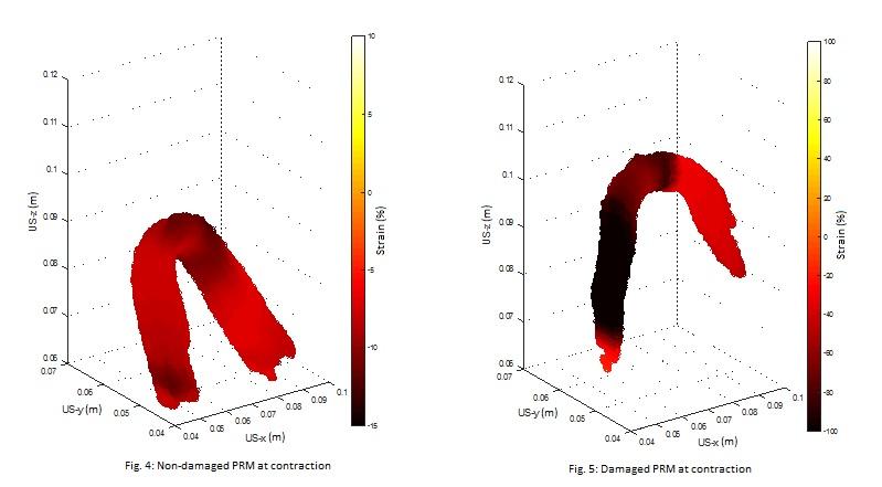
Figure 4-5: Percentage of strain in contracted PRMs (with and without damage)
Funding
- GYNIUS: Gynaecological Imaging using 3D Ultrasound (Dutch Technology Foundation STW and Philips, active)
- Finding Obstetric Complications using UltraSound and Artificial Intelligence (NWO-TTW Demonstrator, active)