Research
BREAST
Background
Breast cancer is a malignant tumour that originates in the cells of the breast. It is one of the most commonly diagnosed types of cancer among women. The lifetime risk of developing breast cancer in industrialized countries is at a level of one in seven women. Early detection and diagnosis are of key importance for effective treatment of breast cancer. However, current diagnostic methods are insufficient to detect all breast cancers in time. This is mainly shown by the condition that we fail to prevent metastatic spread and to adequately monitor treatment. Consequently, new methods are required to improve early cancer detection in a screening setting and after detection to guide and monitor treatment. Ultrasound imaging might be an excellent choice since it has low costs, is real-time en does not use hazardous radiation. However, ultrasound imaging lacks adequate specificity and requires well-educated radiologist to interpret breast ultrasound images. Therefore, our research goal is to improve the detection, diagnosis and monitoring of breast cancer using ultrasound imaging and related methods. Furthermore, we aim to develop low-cost ultrasound and CAD systems to facilitate breast cancer detection for health care workers in developing countries.
3D Doppler and perfusion imaging
One way to improve the specificity of ultrasound imaging is to detect the blood flow in and around suspected lesions. Breast cancers often have higher vascularization due to angiogenesis compared to benign lesions. With ultrasound Doppler techniques and perfusion imaging blood velocities and flows. We are developing 3D Doppler for the automated breast volume scanner to detect low blood flows around lesions. Furthermore, we are investigating ultrasound perfusion imaging using contrast agents (microbubbles) to visualize the vascularisation of lesions and to determine related parameters to classify lesions like wash-in/our curves.
3D ultrafast ultrasound data was obtained from a breast with a malignant lesion using a translating 5 Mhz ultrasound probe which ultrafastly transmitted plane wave ultrasound. The 3D power Doppler volume data is visualized below. Besides the volumetric representation, 2 intersecting planes are presented as slices. It can be observed that few clouds of Doppler signals present at both the center and margin of the lesion. More details on the theory behind the implemented method and simulation results can be found in (Chen, 2021). The presented in vivo images and and additional results have been published in (Chen, 2022)
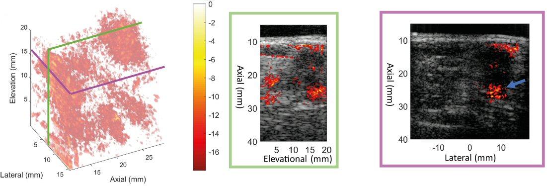
In-vivo experiment on malignant breast lesion. Both Doppler volume (left panel) and two crossing slices (middle and right panel) are presented."