Research
OB-GYN
Abstract Pelvic floor (PF) muscles have the role of preventing pelvic organ descent. The puborectalis muscle (PRM), which is one of the female PF muscles, can be damaged during child delivery. This damage can potentially cause irreversible muscle trauma and even lead to an avulsion, which is disconnection of the muscle from its insertion point, the pubic bone. Ultrasound imaging allows diagnosis of such trauma based on comparison of geometric features of a damaged muscle with the geometric features of a healthy muscle. Although avulsion, which is considered severe damage, can be diagnosed, microdamage within the muscle itself leading to structural changes cannot be diagnosed by visual inspection through imaging only. Therefore, we developed a quantitative ultrasound tissue characterization method to obtain information on the state of the tissue of the PRM and the presence of microdamage in avulsed PRMs. The muscle was segmented as the region of interest (ROI) and further subdivided into six regions of interest (sub-ROIs).

Three-dimensional volume of segmented puborectalis muscle at rest.
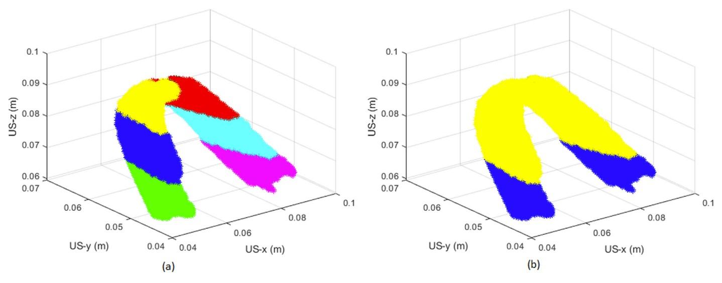
(a) Subregions of interest of puborectalis muscle after division. (b) Two subregions nearest the insertion points at the pubic bone. US = ultrasound.
Mean echogenicity, entropy and shape parameter of the statistical distribution of gray values were analyzed on two of these sub-ROIs nearest to the bone. The regions nearest to the bones are also the most likely regions to exhibit damage in case of disconnection or avulsion. This analysis was performed for both the muscle at rest and the muscle in contraction.
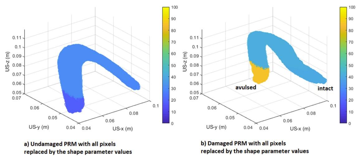
Visual representation of the shape parameter change for (a) an undamaged and (b) a damaged puborectalis muscle (PRM) at rest. US = ultrasound.
We found that, for PRMs with unilateral avulsion compared with undamaged PRMs, the mean echogenicity (p = 0.02) and shape parameter (p < 0.01) were higher, whereas the entropy was lower (p < 0.01). This method might be applicable to quantification of PRM damage within the muscle.