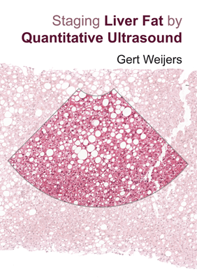
Staging liver fat by quantitative ultrasound
G. Weijers
Promotor Prof. dr. ir. C.L. de Korte, Prof. dr. ir. J.M. Thijssen
Copromotor Dr. G.J.A. Wanten
Institute Radboud University
Date 2019-11-21
In the past decades ultrasound imaging evolved towards an excellent non- invasive imaging technique having high spatial and temporal resolution, from which the parenchymal tissues are characterized by ultrasound texture patterns which can be analyzed qualitatively and quantitatively. Ultrasound imaging has gained a prominent place in clinical practice in studying the liver, since it is noninvasive, radiation free, portable, and relatively cheap compared to other imaging modalities.